-Search query
-Search result
Showing 1 - 50 of 200 items for (author: wagner & t)

EMDB-16538: 
Tomogram of Serratia marcescens in the post-spanin state (1)
Method: electron tomography / : Sitsel O, Wang Z, Janning P, Kroczek L, Wagner T, Raunser S

EMDB-16539: 
Tomogram of Serratia marcescens in the post-spanin state (2)
Method: electron tomography / : Sitsel O, Wang Z, Janning P, Kroczek L, Wagner T, Raunser S

EMDB-16618: 
Structure of Yersinia entomophaga Tc toxin from FIB-milled spheroplasts
Method: subtomogram averaging / : Sitsel O, Wang Z, Janning P, Kroczek L, Wagner T, Raunser S

EMDB-16619: 
Tomogram of Yersinia entomophaga in the post-holin state (whole cell)
Method: electron tomography / : Sitsel O, Wang Z, Janning P, Kroczek L, Wagner T, Raunser S

EMDB-16986: 
Structure of the relaxed thin filament from FIB milled left ventricular mouse myofibrils (tropomyosin masked out)
Method: subtomogram averaging / : Tamborrini D, Wang Z, Wagner T, Tacke S, Stabrin M, Grange M, Kho AL, Rees M, Bennett P, Gautel M, Raunser S

EMDB-16987: 
Structure of the relaxed thin filament from FIB milled left ventricular mouse myofibrils (including tropomyosin)
Method: subtomogram averaging / : Tamborrini D, Wang Z, Wagner T, Tacke S, Stabrin M, Grange M, Kho AL, Rees M, Bennett P, Gautel M, Raunser S

EMDB-16988: 
Tomogram of sarcomere C-zone from mouse cardiac muscle
Method: electron tomography / : Tamborrini D, Wang Z, Wagner T, Tacke S, Stabrin M, Grange M, Kho AL, Rees M, Bennett P, Gautel M, Raunser S

EMDB-16989: 
Tomogram of sarcomere M-band to C-zone from mouse cardiac muscle
Method: electron tomography / : Tamborrini D, Wang Z, Wagner T, Tacke S, Stabrin M, Grange M, Kho AL, Rees M, Bennett P, Gautel M, Raunser S

EMDB-16990: 
Structure of the relaxed thick filament from FIB milled left ventricular mouse myofibrils - Crowns P2-A1
Method: subtomogram averaging / : Tamborrini D, Wang Z, Wagner T, Tacke S, Stabrin M, Grange M, Kho AL, Rees M, Bennett P, Gautel M, Raunser S

EMDB-16991: 
Structure of the relaxed thick filament from FIB milled left ventricular mouse myofibrils - M-band
Method: subtomogram averaging / : Tamborrini D, Wang Z, Wagner T, Tacke S, Stabrin M, Grange M, Kho AL, Rees M, Bennett P, Gautel M, Raunser S

EMDB-16992: 
Structure of the relaxed thick filament from FIB milled left ventricular mouse myofibrils - Crowns A15-A29
Method: subtomogram averaging / : Tamborrini D, Wang Z, Wagner T, Tacke S, Stabrin M, Grange M, Kho AL, Rees M, Bennett P, Gautel M, Raunser S

EMDB-16993: 
Structure of the relaxed thick filament from FIB milled left ventricular mouse myofibrils - Crown P1
Method: subtomogram averaging / : Tamborrini D, Wang Z, Wagner T, Tacke S, Stabrin M, Grange M, Kho AL, Rees M, Bennett P, Gautel M, Raunser S

EMDB-16994: 
Structure of the relaxed thick filament from FIB milled left ventricular mouse myofibrils - Crowns A11-A15
Method: subtomogram averaging / : Tamborrini D, Wang Z, Wagner T, Tacke S, Stabrin M, Grange M, Kho AL, Rees M, Bennett P, Gautel M, Raunser S

EMDB-16995: 
Structure of the relaxed thick filament from FIB milled left ventricular mouse myofibrils - Crowns A8-A12
Method: subtomogram averaging / : Tamborrini D, Wang Z, Wagner T, Tacke S, Stabrin M, Grange M, Kho AL, Rees M, Bennett P, Gautel M, Raunser S

EMDB-16996: 
Structure of the relaxed thick filament from FIB milled left ventricular mouse myofibrils - Crowns A5-A7
Method: subtomogram averaging / : Tamborrini D, Wang Z, Wagner T, Tacke S, Stabrin M, Grange M, Kho AL, Rees M, Bennett P, Gautel M, Raunser S

EMDB-16997: 
Structure of the relaxed thick filament from FIB milled left ventricular mouse myofibrils - Crowns A1-A5
Method: subtomogram averaging / : Tamborrini D, Wang Z, Wagner T, Tacke S, Stabrin M, Grange M, Kho AL, Rees M, Bennett P, Gautel M, Raunser S

EMDB-18146: 
In situ structures from relaxed cardiac myofibrils reveal the organization of the muscle thick filament
Method: subtomogram averaging / : Tamborrini D, Wang Z, Wagner T, Tacke S, Stabrin M, Grange M, Kho AL, Rees M, Bennett P, Gautel M, Raunser S

EMDB-18200: 
Thin filament consensus map from FIB milled relaxed left ventricular mouse myofibrils
Method: subtomogram averaging / : Tamborrini D, Wang Z, Wagner T, Tacke S, Stabrin M, Grange M, Kho AL, Rees M, Bennett P, Gautel M, Raunser S

EMDB-18147: 
Thin filament from FIB milled relaxed left ventricular mouse myofibrils
Method: subtomogram averaging / : Tamborrini D, Wang Z, Wagner T, Tacke S, Stabrin M, Grange M, Kho AL, Bennet P, Rees M, Gautel M, Raunser S

EMDB-18198: 
Helical reconstruction of the relaxed thick filament from FIB milled left ventricular mouse myofibrils
Method: subtomogram averaging / : Tamborrini D, Raunser S
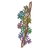
PDB-8q4g: 
Thin filament from FIB milled relaxed left ventricular mouse myofibrils
Method: subtomogram averaging / : Tamborrini D, Wang Z, Wagner T, Tacke S, Stabrin M, Grange M, Kho AL, Bennet P, Rees M, Gautel M, Raunser S

PDB-8q6t: 
Helical reconstruction of the relaxed thick filament from FIB milled left ventricular mouse myofibrils
Method: subtomogram averaging / : Tamborrini D, Raunser S

EMDB-16236: 
Triangle 1 v1 for DNA ORIGAMI TRAPS FOR LARGE VIRUSES
Method: single particle / : Monferrer A, Kohler F, Sigl C, Schachtner M, Peterhoff D, Asbach B, Wagner R, Dietz H

EMDB-16237: 
Triangle-1-version-2 for DNA ORIGAMI TRAPS FOR LARGE VIRUSES
Method: single particle / : Monferrer A, Kohler F, Sigl C, Schachtner M, Peterhoff D, Asbach B, Wagner R, Dietz H

EMDB-16238: 
Triangle-1-version-3 for DNA ORIGAMI TRAPS FOR LARGE VIRUSES
Method: single particle / : Monferrer A, Kohler F, Sigl C, Schachtner M, Peterhoff D, Asbach B, Wagner R, Dietz H

EMDB-16239: 
Triangle-2-version-2 for DNA ORIGAMI TRAPS FOR LARGE VIRUSES
Method: single particle / : Monferrer A, Kohler F, Sigl C, Schachtner M, Peterhoff D, Asbach B, Wagner R, Dietz H

EMDB-16240: 
Triangle-2-version-3 for DNA ORIGAMI TRAPS FOR LARGE VIRUSES
Method: single particle / : Monferrer A, Kohler F, Sigl C, Schachtner M, Peterhoff D, Asbach B, Wagner R, Dietz H

EMDB-16261: 
Triangle-2-version-1 for DNA ORIGAMI TRAPS FOR LARGE VIRUSES
Method: single particle / : Monferrer A, Kohler F, Sigl C, Schachtner M, Peterhoff D, Asbach B, Wagner R, Dietz H

EMDB-16262: 
9-mer cone assembly for DNA ORIGAMI TRAPS FOR LARGE VIRUSES
Method: single particle / : Monferrer A, Kohler F, Sigl C, Schachtner M, Peterhoff D, Asbach B, Wagner R, Dietz H

EMDB-16263: 
10-mer-cone for DNA ORIGAMI TRAPS FOR LARGE VIRUSES
Method: single particle / : Monferrer A, Kohler F, Sigl C, Schachtner M, Peterhoff D, Asbach B, Wagner R, Dietz H

EMDB-15403: 
Structure of the secreted Yersinia entomophaga Tc toxin YenTc
Method: subtomogram averaging / : Sitsel O, Wang Z, Janning P, Kroczek L, Raunser S

EMDB-15404: 
Tomogram of Yersinia entomophaga in the pre-secretory state
Method: electron tomography / : Sitsel O, Wang Z, Janning P, Kroczek L, Raunser S

EMDB-15405: 
Tomogram of Yersinia entomophaga in the post-holin state
Method: electron tomography / : Sitsel O, Wang Z, Janning P, Kroczek L, Raunser S

EMDB-15406: 
Tomogram of Yersinia entomophaga in the post-endolysin state
Method: electron tomography / : Sitsel O, Wang Z, Janning P, Kroczek L, Raunser S

EMDB-15407: 
Tomogram of Yersinia entomophaga in the post-spanin state
Method: electron tomography / : Sitsel O, Wang Z, Janning P, Kroczek L, Raunser S

EMDB-17452: 
Single particle cryo-EM structure of the homohexameric 2-oxoglutarate dehydrogenase OdhA from Corynebacterium glutamicum
Method: single particle / : Yang L, Mechaly AM, Bellinzoni M

EMDB-17453: 
Single particle cryo-EM structure of homohexameric 2-oxoglutarate dehydrogenase OdhA from Corynebacterium glutamicum with Coenzyme A bound to the E2o domain
Method: single particle / : Yang L, Mechaly AM, Bellinzoni M

EMDB-17454: 
Single particle cryo-EM structure of homohexameric 2-oxoglutarate dehydrogenase OdhA from Corynebacterium glutamicum in complex with the product succinyl-CoA
Method: single particle / : Yang L, Mechaly AM, Bellinzoni M

EMDB-17455: 
Single particle cryo-EM structure of homohexameric 2-oxoglutarate dehydrogenase OdhA from Corynebacterium glutamicum following reaction with the 2-oxoglutarate analogue succinyl phosphonate
Method: single particle / : Yang L, Mechaly AM, Bellinzoni M

EMDB-17456: 
Single particle cryo-EM structure of the complex between Corynebacterium glutamicum homohexameric 2-oxoglutarate dehydrogenase OdhA and the FHA-protein inhibitor OdhI
Method: single particle / : Yang L, Mechaly AM, Bellinzoni M

PDB-8p5t: 
Single particle cryo-EM structure of the homohexameric 2-oxoglutarate dehydrogenase OdhA from Corynebacterium glutamicum
Method: single particle / : Yang L, Mechaly AM, Bellinzoni M

PDB-8p5u: 
Single particle cryo-EM structure of homohexameric 2-oxoglutarate dehydrogenase OdhA from Corynebacterium glutamicum with Coenzyme A bound to the E2o domain
Method: single particle / : Yang L, Mechaly AM, Bellinzoni M
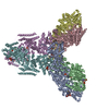
PDB-8p5v: 
Single particle cryo-EM structure of homohexameric 2-oxoglutarate dehydrogenase OdhA from Corynebacterium glutamicum in complex with the product succinyl-CoA
Method: single particle / : Yang L, Mechaly AM, Bellinzoni M
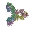
PDB-8p5w: 
Single particle cryo-EM structure of homohexameric 2-oxoglutarate dehydrogenase OdhA from Corynebacterium glutamicum following reaction with the 2-oxoglutarate analogue succinyl phosphonate
Method: single particle / : Yang L, Mechaly AM, Bellinzoni M
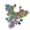
PDB-8p5x: 
Single particle cryo-EM structure of the complex between Corynebacterium glutamicum homohexameric 2-oxoglutarate dehydrogenase OdhA and the FHA-protein inhibitor OdhI
Method: single particle / : Yang L, Mechaly AM, Bellinzoni M

EMDB-29930: 
T. cruzi topoisomerase II alpha bound to dsDNA and the covalent inhibitor CT1
Method: single particle / : Schenk A, Deniston C, Noeske J

PDB-8gcc: 
T. cruzi topoisomerase II alpha bound to dsDNA and the covalent inhibitor CT1
Method: single particle / : Schenk A, Deniston C, Noeske J

EMDB-33247: 
Cryo-EM structure of the Neuromedin U receptor 2 (NMUR2) in complex with G Protein and its endogeneous Peptide-Agonist NMU25
Method: single particle / : Zhao W, Wenru Z, Mu W, Minmin L, Shutian C, Tingting T, Gisela S, Holger W, Albert B, Cuiying Y, Xiaojing C, Han S, Wu B, Zhao Q

PDB-7xk8: 
Cryo-EM structure of the Neuromedin U receptor 2 (NMUR2) in complex with G Protein and its endogeneous Peptide-Agonist NMU25
Method: single particle / : Zhao W, Wenru Z, Mu W, Minmin L, Shutian C, Tingting T, Gisela S, Holger W, Albert B, Cuiying Y, Xiaojing C, Han S, Wu B, Zhao Q

EMDB-27936: 
Cryo-EM structure of Apo form ME3
Method: single particle / : Yu X, Grell TAJ, Shaffer PL, Steele R, Sharma S, Thompson AA, Tresadern G, Ortiz-Meoz RF, Mason M, Gomez-Tamayo JC, Riley D, Wagner MV, Wadia J
Pages:
 Movie
Movie Controller
 Structure viewers
 About EMN search


 wwPDB to switch to version 3 of the EMDB data model
